
prokaryotic cell bacteria parts
Bacterial cells were once presumed to be 'bags of enzymes' with minimal oganization 1. Yet, in the past 10 years, numerous studies have demonstrated that bacteria compartmentalize many.
Cell Structure & Function Notes Mr. Stewart's Biology Class
A fundamental method in molecular biology and microbiology is labeling bacterial cells. Keeping track of and identifying particular bacterial strains, running research, and comprehending bacterial behaviour all depend on it. Here is your comprehensive guide to labeling bacteria cells, with all the information you require: Why Bacterial Cells Are Labeled Monitoring and Recognition: Labels make.
Bacterial Structure Plantlet
1.11: Prokaryotic Cells. Distinguish between prokaryotic cells and eukaryotic cells in terms of structure, size, and the types of organisms that have these cell types. Identify structures of bacterial cells in models and diagrams, including details of Gram-positive and Gram-negative cell walls and flagella.
30 Label A Bacterial Cell
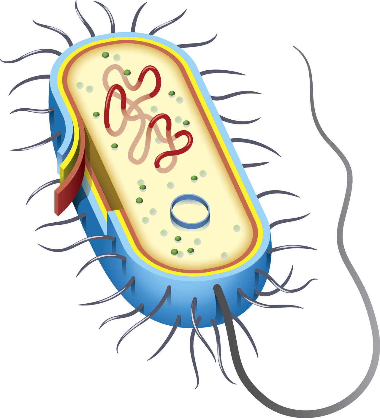
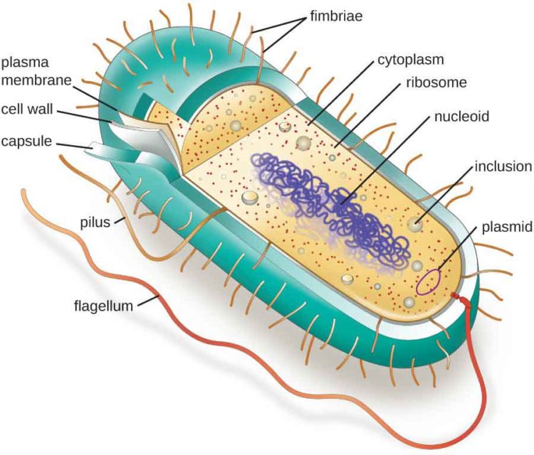
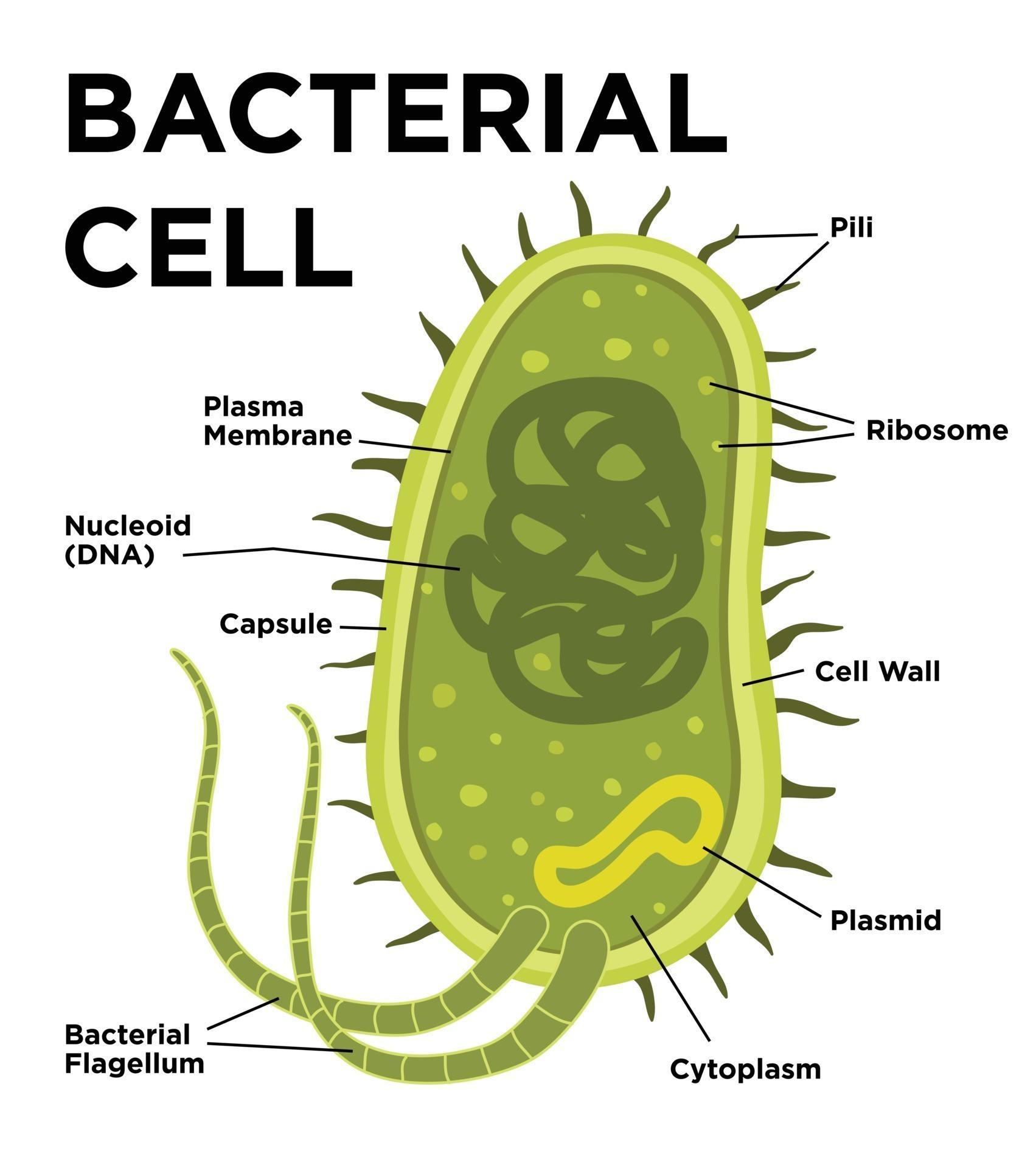
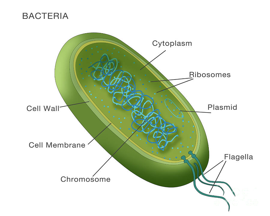
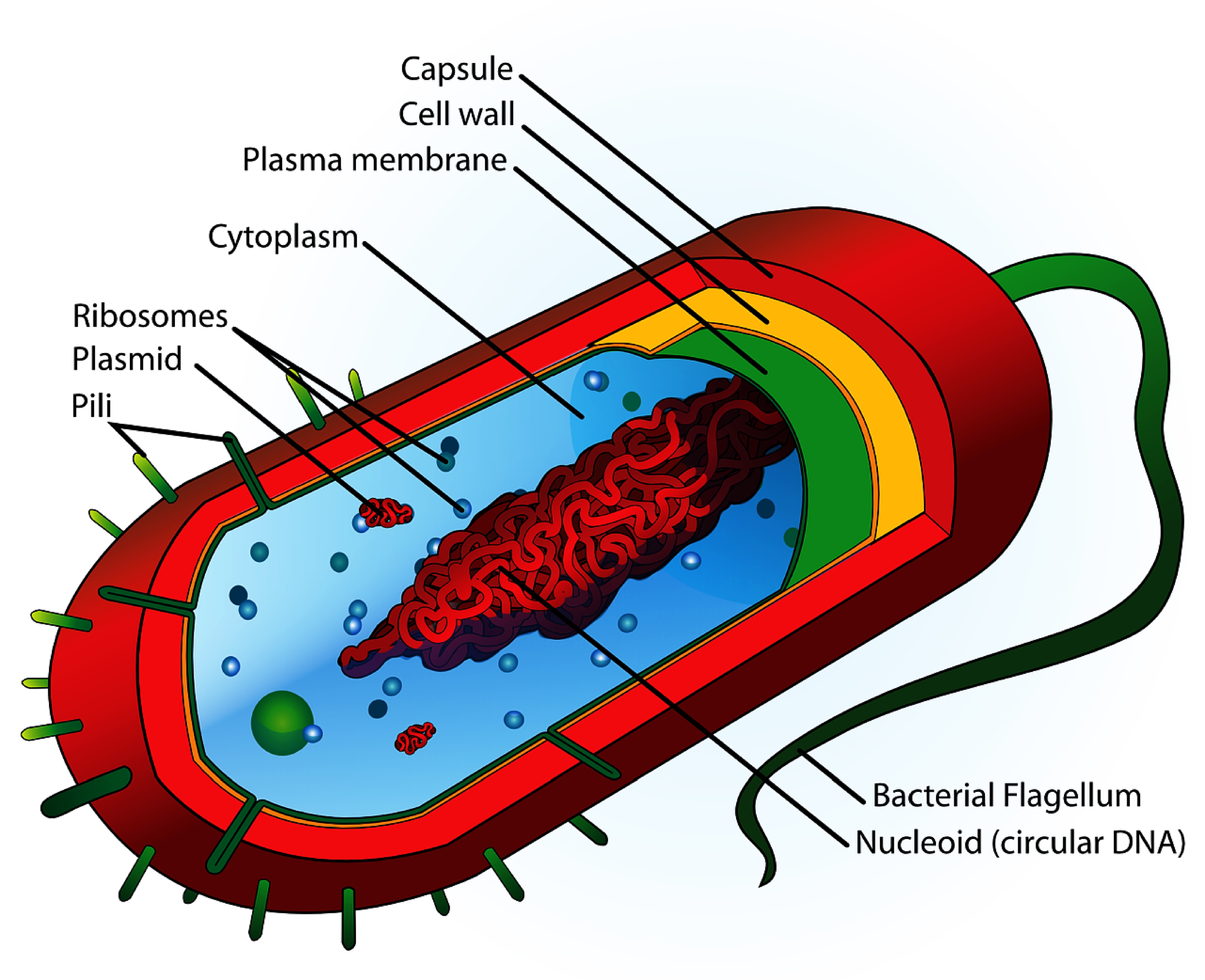
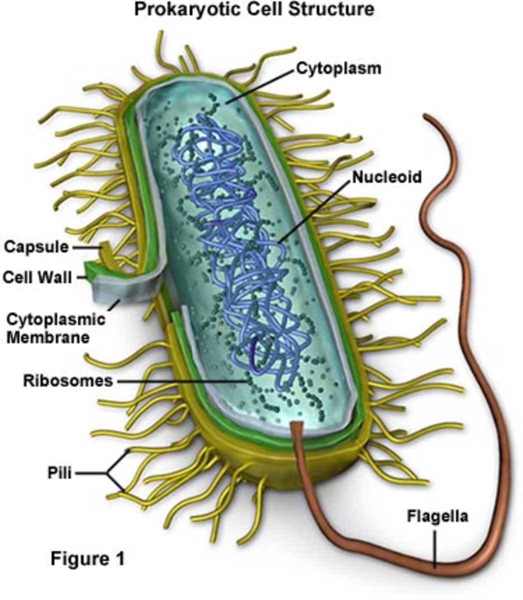
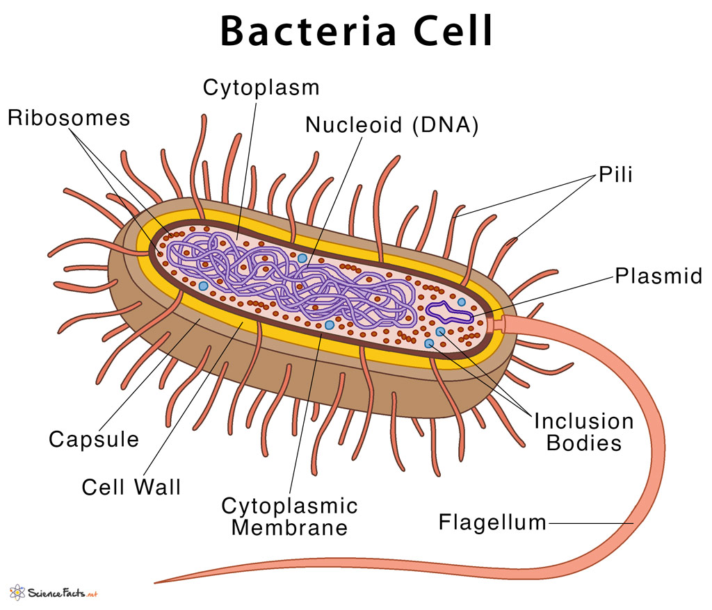
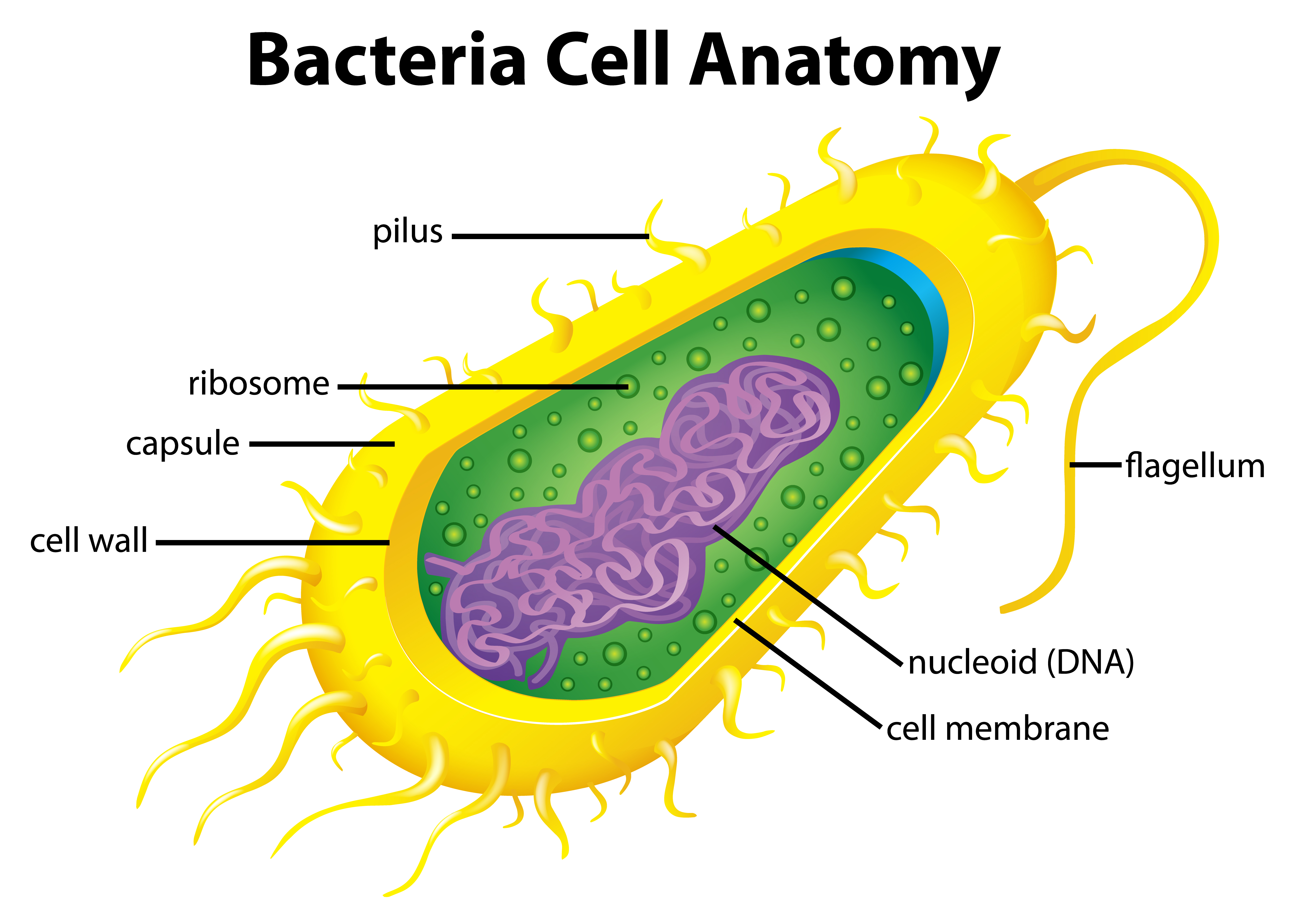
Figure 1. Cutaway drawing of a typical bacterial cell illustrating structural components. See Table 2 below for chemical composition and function of the labeled components. Table 2. Summary of characteristics of typical bacterial cell structures. Structure. Flagella. Function (s) Swimming movement.
Prokaryotic Cell Structure, Characteristics & Function
bacteria, any of a group of microscopic single-celled organisms that live in enormous numbers in almost every environment on Earth, from deep-sea vents to deep below Earth's surface to the digestive tracts of humans. Bacteria lack a membrane-bound nucleus and other internal structures and are therefore ranked among the unicellular life-forms.

Bacteria Structure Over 15,693 RoyaltyFree Licensable Stock Vectors & Vector Art Shutterstock
Prokaryotic cell (bacterial cell) Size: Most are 5 μm - 100 μm: Most are 0.2 μm - 2.0 μm: Outer layers of cell: Cell membrane. Surrounded by cell wall in plants and fungi.
Bacterial Cell Structure and Function
4 Bacteria: Cell Walls . It is important to note that not all bacteria have a cell wall.Having said that though, it is also important to note that most bacteria (about 90%) have a cell wall and they typically have one of two types: a gram positive cell wall or a gram negative cell wall.. The two different cell wall types can be identified in the lab by a differential stain known as the Gram stain.
Bacterial cell anatomy in flat style. Vector modern illustration. Labeling structures on a
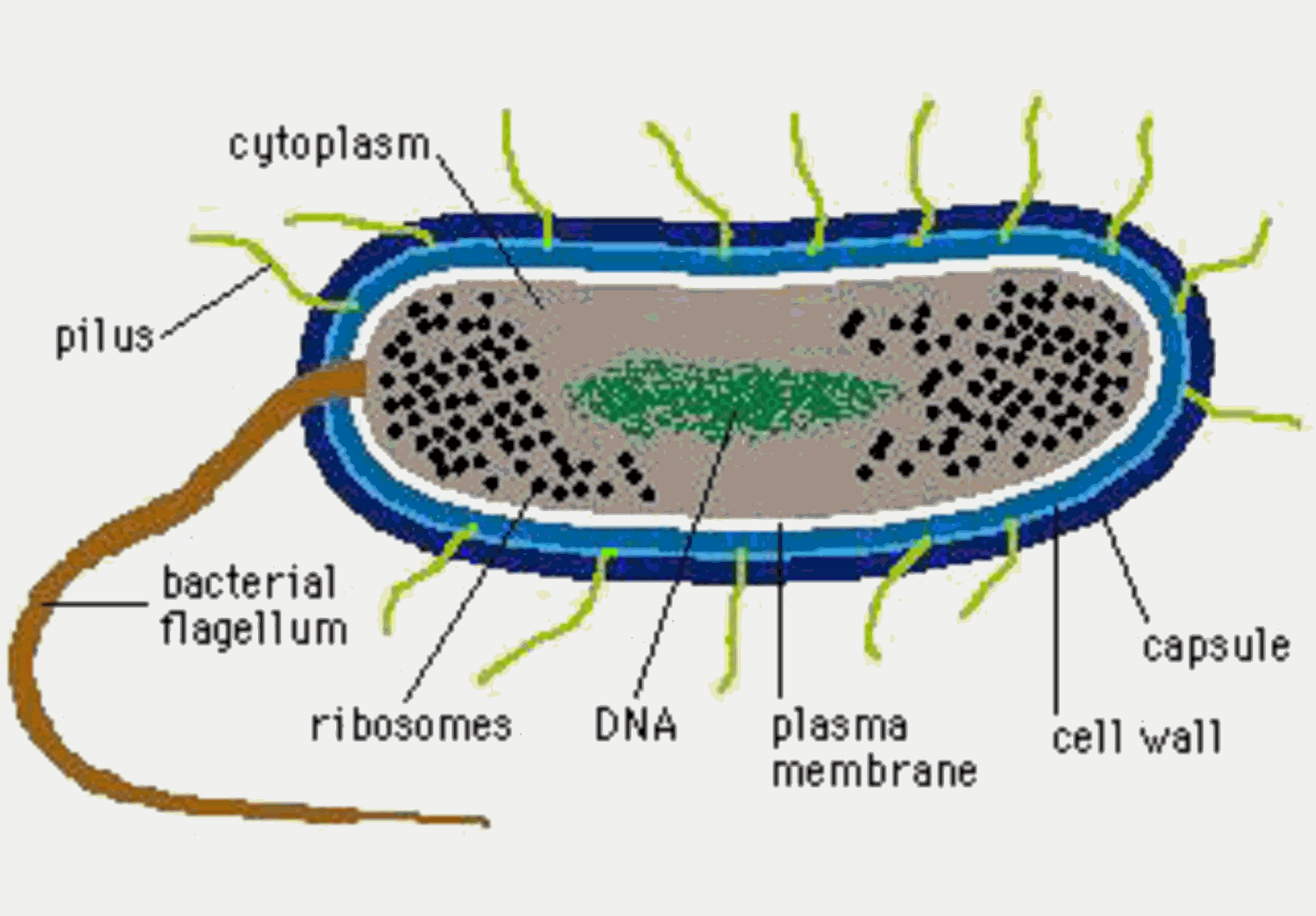
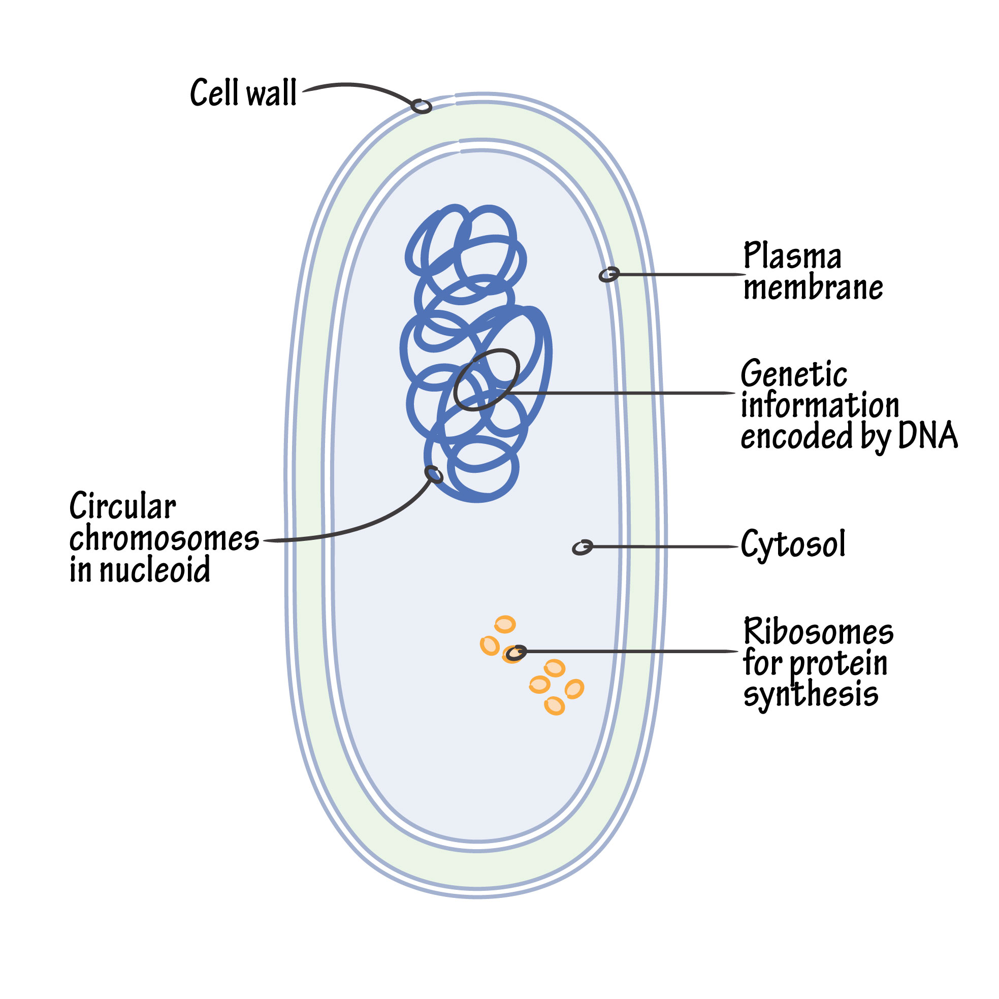
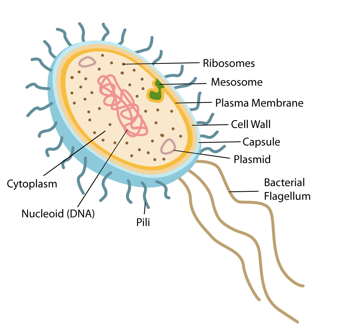
In this article we will discuss about the cell structure of bacteria with the help of diagrams. A bacterial cell (Fig. 2.5) shows a typical prokaryotic structure. The cytoplasm is enclosed by three layers, the outermost slime or capsule, the middle cell wall and inner cell membrane. The major cytoplasmic contents are nucleoid, plasmid, ribosome.
Bacteria Diagram Photograph by Monica Schroeder
In gram-negative bacteria, the cell wall is thin and releases the dye readily when washed with an alcohol or acetone solution. Cytoplasm - The cytoplasm, or protoplasm, of bacterial cells is where the functions for cell growth, metabolism, and replication are carried out. It is a gel-like matrix composed of water, enzymes, nutrients, wastes.

Learn About Prokaryotic Cells, Prokaryotes Bacteria and Archaeans Prokaryotic cell
Cell wall: It is a tough and rigid structure of peptidoglycan with accessory specific materials (e.g. LPS, teichoic acid etc.) surrounding the bacterium like a shell and lies external to the cytoplasmic membrane. It is 10-25 nm in thickness. It gives shape to the cell. Nucleus: The single circular double-stranded chromosome is the bacterial genome.
Effective use of alcohol for aromatic blending Tisserand Institute
Prokaryotes, like bacteria are summarized in Chapter 8. Students learn the difference between prokaryote and eukaryote cells. They learn the basic parts of all cells: membrane, DNA, cytoplasm. You can copy and edit my Google Slides for Chapter 8. Chapter 21 covers bacteria with more details, including the topic with viruses, protists, and fungi.
Bacterial Intracellular Structures That Give Bacteria/Prokaryotes an Advanatage! HubPages
Cell size. Typical prokaryotic cells range from 0.1 to 5.0 micrometers (μm) in diameter and are significantly smaller than eukaryotic cells, which usually have diameters ranging from 10 to 100 μm. The figure below shows the sizes of prokaryotic, bacterial, and eukaryotic, plant and animal, cells as well as other molecules and organisms on a.
Types Of Bacterial Cells
Bacterial cells have simpler internal structures like Pilus (plural Pili), Cytoplasm, Ribosomes, Capsule, Cell Wall, Plasma membrane, Plasmid, Nucleoid, Flagellum, etc. Labeled Bacteria diagram. Eukaryotes have been shown to be more recently evolved than prokaryotic microorganisms. Eukaryotic cells, which make up higher organisms, evolved from.
Bacteria Cell Vector Art, Icons, and Graphics for Free Download
Bacteria cells are the smallest living cells that are known; even though viruses are smaller than bacteria, viruses are not living cells. In microbiology there are different types of bacteria with various sizes, shapes, and structures. The bacteria shapes, structure, and labeled diagrams are discussed below.

Pin by Magpie on ชีวะ Prokaryotic cell, Eukaryotic cell, Prokaryotic cell model
3.3 Bacterial Plasma Membranes. Describe the fluid mosaic model of membrane structure and identify the types of lipids typically found in bacterial membranes. Distinguish macroelements (macronutrients) from micronutrients (trace elements) and provide examples of each. Provide examples of growth factors needed by some microorganisms.

Structure and Function of Prokaryotic Cells
3.1 Cell Culture and Labeling. Depending on the goal of experiments, one can label bacteria with FDAA with a long-pulse, short-pulse, or pulse-chase. In long-pulse labeling, incubate dyes with cell cultures for 1-3 cell cycle durations so that the whole bacterial outline can be visualized; for short-pulse labeling, incubate dyes with cells for 5-10% of the cell cycle so that nascent PG.